Leg muscles, their location, functions and structure. Anterior and posterior muscle groups of the leg
The anatomy of the human lower leg is a complex system of interconnected muscles, bones and ligaments. The development of the muscles of the lower leg determines their structure, as is the case with the muscular apparatus of the thigh or pelvic region - all these areas are responsible for the ability to walk upright, and this type of movement implies high load. The entire muscular complex of the lower leg, intermuscular septa and lower leg fascia (FG) is responsible for the correct functioning of the knees, ankle and foot.
Leg muscles: location, functions
This zone is included in the leg and runs from the knee to the foot. The skeletal foundation of the site is built on only two components - the tibia and the tibia. Musculature covers them in 3 sides. The functionality of the complex:
- implementation of the movement;
- flexion/extension of articular mechanisms.
Tibial segment
It is classified as part of the anterior leg muscle group. This system controls the area of the skeletal apparatus of the considered part of the limb. The tibialis anterior muscle (TBM) begins to develop on the outer plane of the bone bearing the same name. Subsequently, it moves beyond the lower and upper retainers, which unbend the fibers, which are enlarged processes of the leg and foot fascia and develop on the lower leg. Then the PBM is attached to the base of the growth of the first metatarsal, as well as to the medial sphenoid bone.
The muscle is easy to feel through the skin, it is especially noticeable in the place where the foot begins, because. the connective tendon of the fiber protrudes outward. It works as an extensor of the leg muscles and additionally serves as an arch support.
Finger extensor (long)
The DRP is localized on top of the previously mentioned element in the initial segment. Growth starts from the tip of the tibia and the frontal marginal surface of the fibula, from the FG and the interosseous membrane. At the foot level, the fiber is separated into 5 tendons (SG):
- 4 are attached to toes 2 to 5;
- the last - to the beginning of the 5th metatarsal bone.
The long extensor of the fingers performs a function understandable from the name also for the foot. Due to the tendon attachment to the outer side of the foot, the element also has the ability to pronate.
Extensors of the thumbs
Between the middle of the PBM and the sidewall of the DRP, which is sometimes covered in the anterior region by these muscles, there is a long extensor thumb. It is formed in the second third of the frontal surface of the fibula and the joints of the elements of the lower leg. The tendons belonging to the muscle move towards the heel, spreading behind the holders mentioned above in a separate synovial sheath, after which they join the distal phalanx of the thumb as a whole, and optionally to the next one after the nail. The task of the muscle of the anterior surface of the lower leg is to straighten the BP and provide motor ability foot area in the ankle.
Finger flexor
DSP (long flexor of the fingers) departs from the back of the tibia and moves towards the sole, slipping behind the medial malleolus in a special channel lying below the fixator.
Near the plantar surface, Digitorum longus runs through the tendon that flexes the big toe, a square muscle is attached to it, which subsequently disperses into 4 striated muscles connected to the DF (distal phalanges) from the second to 5 fingers.
The element supinates the foot and makes the toes clench. The task of the square muscle is to balance the impact, which is necessary, because. the divided part of the chipboard performs flexion, and also balances the limb to the median plane of the body. The attached muscular structure pulls outward, the adductor action weakens, and flexion is carried out rather in the vertical plane of the body.

Triceps muscle of the leg
Belongs to the muscles rear surface shins. The name is due to its structure, because. has three muscle ends (heads):
- the first and second are closer to the dermis and form calves;
- the third lies deeper in the limbs and makes up the soleus muscle, holds the site on the talus, without moving forward.
The processes are connected to the Achilles tendon, fastened to the tubercle of the calcaneus.
The medial and lateral condyles of the femoral region are the starting point for the growth of the calves. The second head is less developed than the first, descending a little further. They have two bending tasks:
- in the knee;
- ankle joint.
The soleus head grows from the back of the upper third of the BB bone and from the tendon between the tibial and fibular parts of the skeleton. Located behind the subtalar joint and ankle, the fiber regulates the fold of the medial edge of the foot.
In the superficial visible part, the triceps muscle of the lower leg stands out visually and is examined by touch without difficulty. Characterized by a maximum range of rotation perpendicular to the ankle joint due to the fact that the heel ligaments in the rear of the foot stand out behind the mentioned axis.
The diamond-shaped popliteal fossa is formed by the heads of the gastrocnemius muscle. The rhombus limits the posterior muscle group of the lower leg, as well as:
- Anterior part - biceps femoral area.
- Back and top - semimembranosus muscle.
- In the lower part - the plantar muscle and the ends of the calf.
- Bottom - capsule knee joint and thigh.
Threads of nerve endings and arteries are laid along the bottom, feeding and controlling muscle and bone tissue.
thumb flexor
The most powerful muscle in the lower leg - hallucis longus - develops from the bottom of the rear section of the MBC and the back membrane. Near the sole, the muscle lies in the middle of the components of the BP flexor minor, grows from the beginning of the distal phalanx of the first toe.

The purpose of the existence of the tendon of the long flexor of the large, or first, finger in the body is to squeeze it and the foot.
Due to partial fusion with the tendon of the flexor process, the position of the second and third fingers is affected. There are 2 sesamoid bones near the metatarsophalangeal joint of the BP, thanks to which the moment of rotation of the DSBP increases.
Tibialis posterior
It is localized deeper than the triceps between the flexor muscles of the lower leg. Beginning - the back side of the interosseous septum and closely lying parts of the tibia. After passing through the medial malleolus, the muscle is attached to the tubercle of the navicular and sphenoid bones, to the metatarsus. The tibialis posterior muscle, which belongs to the adductor muscles of the lower leg, is responsible for the following actions:
- bringing the foot into motion;
- supination;
- bending.
The canal separates the fiber from the soleus muscle, the so-called. calf-popliteal, from the front resembling a thin slit. In its bed lie nerve fibers and blood vessels.
Second division of tibial fibers
It begins to form in the same place as the muscle described in the paragraph earlier, and is placed in the mass of tissues, in contrast to the triceps. It is attached to the metatarsal, sphenoid and scaphoid bones. This fragment of the lateral muscle group of the lower leg, combined with the MBBM, bends and moves the foot.
Popliteal segment
Consists of a complex of connected small fibers lying near the surface of the knee. They pass:
- from the lateral condyle of the thigh;
- deeper than the calf area and knee synovial bursa;
- rise above the soleus muscle and are attached to the tibia.
Since the muscle strips are partially attached to the knee bag, during flexion, the bursa is pulled back.
The functional tasks performed by the popliteal muscle include:
- ensuring the mobility of the lower leg;
- her natural pronation.
Long peroneal segment

A distinctive feature of the site is its pinnate structure. The muscle lies on top of the MB of the bone, is attached to its 2 thirds from the outer part, growing from:
- its head part;
- partially - fascia;
- condyle BBK.
When the long peroneal muscle contracts, 3 types of movement are immediately provided:
- abduction;
- pronation (bend);
- the leg is bent at the foot.
The tendon of this fiber wraps around the lateral part of the ankle behind and below. Near the heel they meet the extreme retainers. Moving further and getting surrounded by the muscles of the sole, the element spreads along the groove running along the lower surface of the cuboid bone of the foot, and ends on its inner side.
Short peroneal fibers
It is this subtype of flat muscle formations that raises the lateral edge of the foot, does not allow it to turn with the plantar side inward and clubfoot, and performs plantar flexion.
A short MB fiber is formed by the fusion of the shin septa and the fibula on its superficial side facing the skin. As it moves down and isolates the tendon from the short peroneal muscle, it fits the ankle lateral structure from the rear lower edge, after which it is attached to the tuberous protrusion of the last metatarsal bone.
Common malformations

In addition to serious but rare anomalies, such as the absence of one of the limbs or some of their parts, fusion together and other global defects, among the pathologies of the formation of bones and muscles of the lower leg, there are:
- Curvature of the leg in the frontal plane - can go away by itself after the baby learns to walk independently, and treatment is not required.
- Native subluxation or dislocation is often bilateral, its companion is a change in the shape of the knees and contracture. The diagnosed type of deformation depends on the strength and nature of the changes. The changes are due to the fact that the muscles are attached in the wrong places, due to the underdevelopment of the femur and tibia. Such a pathology may be accompanied by problems with the structure and function of the ankle, underdevelopment or complete absence of the tibia.
- Hypoplasia (underdevelopment and small size) of the elements.
- The presence of false joints, constriction by ligaments of supply nodes.
Even with proper development leg structures may develop abnormalities as they grow, caused by a lack of bone mineralization, inflammation in the joints and muscles, excessive or insufficient loads, injuries, improper selection of shoes or malnutrition.
The lower leg is a complex structure, consisting of many finely adjusted components, so this part of the body can be subject to pathological changes. A high permanent load increases the risk of developing diseases and defective conditions. It needs to be given attention in general health care, especially in infants on early stages post-natal development and in the elderly due to the vulnerability of the joints and the fragility of the bone tissue. When eating, it is necessary to maintain the level of useful microelements for the human skeleton, periodically taking a complex of vitamins. It is also necessary to monitor the condition of the joints, if possible, reduce the load on the limbs with the help of specialized orthopedic devices and develop muscles.
The shin belongs to lower limb. It is located between the foot and the knee area. The lower leg is formed by means of two bones - small and tibial. They are surrounded by muscle fibers on three sides. The muscles of the lower leg, the anatomy of which will be discussed later, set the fingers and foot in motion.
Tibia
This element has an extension on the top edge. Condyles are formed in this area: lateral and medial. On top of them are the surfaces of the joints. They articulate with the condyles of the thigh. On the lateral segment, there is an articular surface on the outside, through which it is connected to the head in the fibula. The body of the tibial element looks like a trihedral prism. Its base is directed backwards and has 3 surfaces, respectively: back, outer and inner. There is an edge between the last two. It's called the front. In its upper part, it passes into the tuberosity of the tibia. This area is intended for fixation. In the lower part, the tibia has an extension, and there is a protrusion on the inner surface. It is oriented downward. This protrusion is called the medial malleolus. On the back side of the bone lies a rough segment of the soleus muscle. The articular surface is located on the distal epiphysis. It serves to connect with

Second element
The fibula is thin, long, located laterally. Its upper end has a thickening - the head. It connects to the tibia. The lower section of the element is also thickened and forms the lateral malleolus. She, like the head of the fibula, is oriented outward and is well palpable.
Leg muscles: their location, functions
The fibers are located on three sides. Allocate different muscles shins. The front group performs extension of the foot and fingers, supination and adduction of the foot. This segment includes three types of fibers. The tibialis anterior muscle of the lower leg was formed first. The remaining fibers form the long extensors of the fingers and a separate one for the big toe on the foot. The posterior muscle group of the lower leg forms a greater number of fibers. In particular, there are long finger flexors and separately for the large, popliteal, triceps muscle of the lower leg. There are also tibial fibers here. The outer group includes the short and long peroneal muscles of the lower leg. These fibers flex, penetrate, and abduct the foot.
Tibial segment
This anterior muscle of the lower leg starts from the bone of the same name, its outer surface, fascia and interosseous membrane. They are directed downward. The fibers pass under two ligaments. They are located in the area and ankles. These areas - the upper and lower retainers of the extensor tendons - are represented by places of thickening of the fascia of the foot and lower leg. The site of attachment of the fibers is the sphenoid medial and the base of the metatarsal (first) bone. The muscle is quite well palpable along its entire length, especially in the area of transition to the foot. In this place, her tendon protrudes during extension. The task of this leg muscle is the supination of the foot.
Finger extensor (long)
It runs from the anterior muscle outward to upper area shins. Its fibers begin from the head and marginal sections of the tibia, fascia and interosseous membrane. The extensor, passing to the foot, is divided into five tendons. Four are attached to the distal (from the second to the fifth), the last - to the base of the 5th metatarsal. The task of the extensor, acting as a multi-joint muscle of the lower leg, is not only to coordinate the extension of the fingers, but also the foot. Due to the fact that one tendon is fixed at its edge, the fibers also penetrate the area somewhat.
Extensors of the thumbs
The fibers begin in the region of the lower leg from the interosseous membrane and the inner part of the fibula. The extensors have less strength than the segments described above. The site of attachment of this is the distal phalanges in the thumbs. These muscles of the lower leg not only carry out their extension, but also the feet, also contributing to their supination.

Finger flexor (long)
It starts from the back of the tibia, passing under the medial malleolus to the foot. The channel for it is located under the retainer. Next, the muscle is divided into four segments. On the foot (plantar surface), fibers cross the tendon from the flexor (long) thumb. Then the square muscle of the sole joins them. Four formed tendons are fixed to the distal phalanges (at their base) of 2-5 fingers. The task of this muscle is, among other things, to flex and supinate the foot. The fibers of the square segment are attached to the tendon. Due to this, the action of the muscle is averaged. Lying under the medial malleolus and fanning out towards the phalanges, the long flexor also provokes some adduction of the fingers to the median surface of the body. By pulling the square muscle of the tendon, this action is slightly reduced.
Triceps muscle of the leg
It runs along the back surface and has 3 heads. Two form the surface area - the gastrocnemius muscle, from the third - deep - the fibers of the soleus segment depart. All heads are connected and form a common Achilles (calcaneal) tendon. It is attached to the tubercle of the corresponding bone. The gastrocnemius muscle starts from the femoral condyles: lateral and medial. The task of the two heads located in this area is twofold. They coordinate flexion at the knee joint and the foot at the ankle joint. The medial element descends slightly lower and is better developed than the lateral one. From the back side in the upper third of the tibia, the soleus muscle departs. It is also attached to the tendon arch located between the bones. The fibers pass somewhat lower and deeper than the gastrocnemius. They lie behind the subtalar and cause flexion of the foot. The triceps muscle can be felt under the skin. From the transverse axis to ankle joint posteriorly protrudes the calcaneal tendon. Due to this, the triceps muscle has a large moment of rotation relative to this line. The heads of the gastrocnemius segment are involved in the formation of the rhomboid popliteal fossa. As its boundaries are: the biceps femoral muscle (outside and top), semimembranous fibers (inside and top), plantar and two heads of the gastrocnemius segment (bottom). The bottom in the fossa is formed by the capsule of the knee joint and the vessels and nerves that feed the foot and lower leg run through this area.

Flexor (long) thumb
This muscle of the posterior surface of the lower leg is characterized by the greatest strength. On the plantar side of the foot, fibers run between the heads from a short segment responsible for flexion of the big toe. The muscle starts from the back side (lower part) of the fibula and the intermuscular septum (back). The site of fixation is the plantar surface of the base of the distal phalanx in the thumb. Due to the fact that the tendon of the muscle partially passes into the element of the long flexor of the same name, it has some influence on the movements of 2-3 fingers. The presence on the surface of the sole of the metatarsophalangeal joint of 2 large sesamoid bone elements provides an increase in the moment of rotation of the fibers. The tasks of the segment include flexion of the entire foot and thumb.
Second division of tibial fibers
This posterior segment is located under the triceps muscle. The fibers start from the interosseous membrane and areas of the small and tibial bones adjacent to it. The site of attachment of the muscle is the tubercle of the navicular, the base of the metatarsal and all the wedge-shaped elements. The muscle lies under the medial malleolus and performs flexion of the foot, supination and adduction. A canal passes between the soleus and tibial fibers. It is presented in the form of a gap. It contains nerves and blood vessels.

Popliteal segment
It is formed by flat short fibers. The muscle adjoins directly to the knee joint from behind. The fibers originate from the femoral condyle (lateral), below the gastrocnemius segment, and the bursa of the knee joint. They pass down and are attached above the soleus muscle to the tibia. Because the fibers are partially attached to the joint capsule, they pull it posteriorly when flexed. The task of the muscle is pronation and flexion of the lower leg.
Long peroneal segment
This muscle has a feathery structure. It runs along the surface of the fibula. It starts from its head, the condyle of the tibial element, partly from the fascia. It is also attached to the 2-thirds region. outside fibula. When the muscle contracts, abduction, pronation, and flexion of the foot occur. The tendon of the long peroneal segment posteriorly and inferiorly bypasses the lateral malleolus. In the area of the heel bone there are ligaments - the upper and lower retainers. When moving to the plantar part of the foot, the tendon runs along the groove. It is located on the underside of the cuboid bone. The muscle reaches the inside of the foot.

Short peroneal fibers
The tendon of the segment wraps around the lateral malleolus behind and below. It is attached to the tubercle on the 5th metatarsal. The segment begins from the intermuscular septa and the outer part of the fibula. The task of the fibers is abduction, pronation and flexion of the foot.
Anterior tibial muscle (m.tibialis anterior) is located on the front side of the lower leg. It begins on the lateral condyle and the upper half of the lateral surface of the body of the tibia, as well as the adjacent part of the interosseous membrane and on the fascia of the leg. At the level of the distal third of the lower leg, the muscle bundles pass into a long tendon that runs under the upper and lower extensor tendon retinaculum, anterior to the ankle joint. Further, the tendon goes around the medial edge of the foot and is attached to the plantar surface of the medial sphenoid bone and the base of the first metatarsal bone.
Function: unbends the foot in the ankle joint, simultaneously raises the medial edge of the foot and turns it outward (supination), strengthens the longitudinal arch of the foot. With a fixed foot, tilts the lower leg forward; helps to keep the leg in a vertical position.
Blood supply: anterior tibial artery
The long extensor digitorum longus (m.extensor digitorum longus) is a pinnate muscle, it begins on the lateral condyle of the tibia, the anterior surface of the body of the fibula, on the upper third of the interosseous membrane, fascia and anterior intermuscular septum of the leg. Heading to the rear of the foot, the muscle passes behind the upper and lower extensor tendon retainers. At the level of the ankle joint, the muscle is divided into 4 tendons, which are enclosed in a synovial sheath common to them. Each tendon is attached to the back of the base of the middle and distal phalanges of the II-V fingers.
A small bundle is separated from the lower part of the muscle, called third peroneal muscle(m.peroneus tertius), the tendon of which is attached to the base of the fifth metatarsal bone.
Function: unbends the II-V fingers in the metatarsophalangeal joints, as well as the foot in the ankle joint. The third peroneal muscle raises the lateral edge of the foot. With a strengthened foot, the long extensor of the fingers holds the lower leg in a vertical position.

Innervation: deep peroneal nerve (LIV-SI). Blood supply: anterior tibial artery.
The long extensor of the big toe (m.extensor hallucis longus) is located between the anterior tibial muscle medially and the long extensor of the fingers laterally; partially covered by them in front. It starts on the middle third of the anterior surface of the fibula, the interosseous membrane of the leg. The tendon of the muscle passes down the dorsum of the foot under the superior and inferior extensor tendon retinaculum in a separate synovial sheath and inserts on the distal phalanx of the big toe. Separate tendon bundles can also attach to the proximal phalanx.
Function: unbends the big toe; also involved in the extension of the foot in the ankle joint.
Innervation: deep peroneal nerve (LIV-SI).
Blood supply: anterior tibial artery.
, , ,
Posterior leg muscles
The muscles of the posterior group form two layers - superficial and deep. The superficially lying triceps muscle of the lower leg is more strongly developed, which creates the roundness of the lower leg characteristic of a person. deep layer It is formed by a small popliteal muscle and 3 long muscles: the long flexor of the fingers (located most medially), the posterior tibial muscle (occupies an intermediate position) and the long flexor of the big toe (located laterally).

Superficial layer of the posterior leg muscles
The triceps muscle of the lower leg (m.triceps surae) consists of two muscles - the gastrocnemius muscle, which is located superficially, and the soleus muscle, hidden under the gastrocnemius. The gastrocnemius muscle is a biarticular muscle, it acts on two joints - the knee and ankle, while the soleus muscle is single-joint - it acts only on the ankle joint.
Calf muscle(m.gastrocnemius) has two heads: medial and lateral, the surface layers of which are represented by strong tendon bundles. The lateral head (caput laterale) begins on the outer surface of the lower epiphysis of the thigh above the lateral condyle. The medial head (caput mediate) begins on the medial condyle of the thigh. Under each head of the gastrocnemius muscle is a synovial bag. Between the lateral head and the capsule of the knee joint is located lateral tendon bursa of the gastrocnemius muscle(bursa subtendinea musculi gastrocnemii lateralis). Between the medial head and the joint capsule is medial gastrocnemius bursa(bursa subtendinea musculi gastrocnemii medialis). Both bags, as a rule, communicate with the cavity of the knee joint.
In the middle of the lower leg, both heads of the gastrocnemius muscle pass into a thick tendon, which tapers downward and merges with the tendon of the soleus muscle, forming the calcaneal (Achilles) tendon (tendo calcaneus, s.Achilli), which is attached to the calcaneal tuberosity. Between the tendon and the calcaneus there is a bag of the calcaneal (Achilles) tendon (bursa tendinis calcanei, s.Achillis).
soleus muscle(m.soleus) thick, flat, lies under the calf muscle. In front of it are the muscles of the deep layer. The soleus muscle has an extensive origin on the posterior surface of the tibia (on the line of the soleus muscle) and on the tendon arch (arcus tendineus musculi solei), which extends between the tibia and fibula. The soleus muscle has a pinnate structure, passes into a flat tendon, which is involved in the formation of the calcaneal tendon.
Function: triceps flexes the lower leg and foot (plantar flexion); with a fixed foot, it holds the lower leg on the talus, preventing it from tipping forward.
Innervation: tibial nerve (LIV-SI).
plantar muscle
(m.plantaris) fickle, has a small abdomen and a long thin tendon. It originates on the lateral epicondyle of the thigh and on the oblique popliteal ligament. The tendon of this muscle passes between the gastrocnemius and soleus muscles, is adjacent to the medial edge of the calcaneal tendon, with which it is attached to the calcaneal tuberosity.
Function: stretches the capsule of the knee joint, participates in flexion of the lower leg and foot.
Deep layer of the posterior leg muscles
The deep layer is formed by 4 muscles: the popliteal, the long flexor of the fingers, the long flexor of the big toe and the posterior tibial muscle, which are separated from the soleus muscle by the deep plate of the fascia of the lower leg.

The popliteal muscle (m.popliteus) lies deep in the popliteal fossa. It begins with a thick tendon on the outer surface of the lateral condyle of the thigh (below the attachment of the peroneal collateral ligament). The muscle is adjacent to the posterior surface of the joint capsule and is located below the arcuate popliteal ligament, on which its medial bundles begin. The muscle attaches to a triangular area on the posterior surface of the tibia, above the line of the soleus muscle.
Function: flexes the lower leg, turning it inwards; stretches the capsule of the knee joint, protecting the synovial membrane from infringement.
Innervation: tibial nerve (LIV-SII).
Blood supply: popliteal artery.

The long flexor of the fingers (m.flexor digitorum longus) has a two-pinnate structure, begins with fleshy bundles on the posterior surface of the body of the tibia below the line of the soleus muscle, as well as on the fascia and posterior intermuscular septum of the leg. It is located behind and medial to the posterior tibial muscle. The tendon of the long flexor of the fingers goes down, crosses behind and from the lateral side the tendon of the posterior tibial muscle. Further, the tendon of the muscle passes to the sole of the foot behind the medial malleolus under the retinaculum of the flexor tendons in a separate synovial sheath (between the tendons of the posterior tibial muscle medially and the long flexor of the thumb laterally). Then the tendon goes around behind and below the support of the talus. Located above the short flexor of the fingers, it is divided into 4 separate tendons, which are attached to the distal phalanges of the II-V fingers, having previously pierced the tendons of the short flexor of the fingers (like the tendons of the deep flexor of the fingers on the hand).
Function: bends the distal phalanges of the II-V fingers; flexes the foot, turning it outward.
Innervation: tibial nerve (LIV-SII).
Blood supply: posterior tibial artery.
Long flexor of the big toe
(m.flexor hallucus longus) - bipennate muscle, begins on the lower two-thirds of the body of the fibula, interosseous membrane, posterior intermuscular septum of the leg. It is located laterally and behind the tibialis posterior muscle. The flexor hallucis longus tendon passes under the flexor tendon retinaculum behind the medial malleolus and lateral to the flexor hallucis longus tendon in a separate synovial sheath. Further, the tendon of the long flexor of the big toe lies in the groove of the same name on the posterior process of the talus, passing forward under the support of the talus. Having reached the plantar surface of the big toe, the tendon of the long flexor of the big toe is attached to its distal phalanx. On its way on the foot, this tendon crosses with the tendon of the long flexor of the fingers (lies under it). Throughout the plantar surface of the I metatarsal bone, the tendon of the long flexor of the big toe lies between the medial and lateral bellies of the short flexor of the big toe.
Function: flexes the big toe, participates in flexion (supination) and adduction of the foot; strengthens the longitudinal arch of the foot.
Innervation: tibial nerve (LIV-SII).
Blood supply: posterior tibial and peroneal arteries.
The posterior tibialis muscle (m.tibialis posterior) is located deep on the back of the leg between the long flexor of the fingers (medially) and the long flexor of the big toe (laterally). It begins on the posterior surface of the body of the fibula (between the medial crest and interosseous margin), the inferior surface of the lateral condyle, and on the upper two-thirds of the body of the tibia (below the line of the soleus muscle) and the interosseous membrane of the leg.
The muscle continues into a strong tendon that lies in a groove on the posterior surface of the medial malleolus in front of the tendon of the long flexor of the fingers (under the retinaculum of the flexor tendons). Moving to the plantar surface of the foot, the tendon is attached to the tuberosity of the navicular bone, to all 3 cuneiform bones, and also to the base of the IV (sometimes V) metatarsal bone.
Function: flexes the foot (plantar flexion), adducts the foot and supinates it.
Innervation: tibial nerve (LIV-SII).
Blood supply: posterior tibial artery.
Lateral leg muscle group
The lateral group is represented by a long and a short peroneal muscles, which are located on the lateral surface of the lower leg under the fascia between the anterior and posterior intermuscular septa.
The long peroneal muscle (m.peroneus longus) is two-pinnate, lies superficially, begins on the head and upper two-thirds of the lateral surface of the fibula, on the lateral condyle of the tibia, the fascia of the lower leg and on the intermuscular septa of the lower leg. At the level of the ankle joint, the tendon of the muscle, bending around the lateral ankle from behind, passes first under the upper retinaculum of the tendons of the peroneal muscles in the common synovial sheath with the tendon of the short peroneal muscle, and then in the groove on the calcaneus (under the lower retinaculum of the tendons of the peroneal muscles). On the sole, the tendon of the long peroneal muscle runs obliquely forward and medially, lies in the groove of the same name in the cuboid bone in a separate (own) synovial sheath. The tendon is attached to the base of the I and II metatarsal bones and to the medial sphenoid bone.

At points where the tendon changes direction (behind the lateral malleolus and on the cuboid bone), it usually thickens due to the fibrocartilage or sesamoid bone that forms in its thickness.
Function: flexes the foot, raises its lateral edge (pronation), strengthens the transverse and longitudinal arches of the foot.
Blood supply: lateral inferior genicular artery, peroneal artery.
The short peroneal muscle (m.peroneus brevis) is two-pinnate, begins on the lower two-thirds of the lateral surface of the fibula and on the intermuscular septa of the leg. The tendon of the muscle passes to the foot behind the lateral ankle under the tendon retinaculum of the peroneal muscles, lying in the common synovial sheath along with the tendon of the long peroneal muscle. At the lower edge of this retainer, the tendon of the short peroneal muscle turns forward and passes along the outer side of the calcaneus under the fibular block to the place of attachment at the base of the fifth metatarsal bone.
Function: raises the lateral edge of the foot; prevents the foot from turning with the sole inside; flexes the foot (plantar flexion).
Innervation: superficial peroneal nerve (LIV-SI).
Blood supply: peroneal artery.
Anterior leg muscles
Anterior tibial muscle (m. tibialis anterior) (Fig. 197) is located on the anterior surface of the lower leg. It has a wide origin from the lateral upper third of the tibia, the fascia of the lower leg and the interosseous membrane. It passes near the anterior tibial crest under the retinaculum mm. extensorum superius et inferius in the fibrous canal and exits on the medial edge of the foot, where the tendon is attached to the plantar surface of the I sphenoid and metatarsal bones.
Function. Extends at the ankle joint and supinates the foot.
The long extensor of the first finger (m. extensor hallucis longus) (Fig. 197) is located lateral to m. tibialis anterior. It starts from the fibula and the interosseous membrane. It comes out between the anterior tibial muscle and the long extensor of the fingers. The tendon passes through the fibrous channel under the retinaculum mm. extensorum superius et inferius, ends at the base of the distal phalanx of the first finger.
Innervation: n. peroneus profundus (LIV-SI).
Function. Corresponds to the name of the muscle. In addition, the muscle is involved in the extension of the foot in the ankle joint.
197. Muscles of the lower leg and foot. 1 - tendo m. sartorius; 2 - tibia; 3 - m. gastrocnemius; 4 - m. soleus; 5 - m. tibialis anterior; 6 - tendo m. extensoris hallucis longi; 7 - tendo m. extensoris digitrum longi; 8 - retinaculum mm. extens6rum inferius; 9 - m. peroneus brevis; 10 - m. peroneus longus; 11-lig. patellae; 12 - tractus iliotibialis.
The long extensor of the fingers (m. extensor digitorum longus) is located lateral to m. tibialis anterior, covers the long extensor of the first finger. It starts from the upper third of the tibia, fibula, membrana interossea and fascia of the leg. The muscle is delimited from the anterior tibial muscle by the intermuscular septum. Forms a tendon that runs in the fibrous sheath under the retinaculum mm. extensorum inferius. Upon reaching the foot, the tendon is divided into 4 tendons, which are attached to the aponeurotic plate of the rear of the II-V fingers.
Innervation: n. peroneus profundus (LIV-SI).
Function. Unbends fingers II-IV, penetrates the outer edge of the foot together with the third peroneal muscle.
The third peroneal muscle (m. peroneus tertius) represents the fifth part of the long extensor of the fingers. This muscle is unstable (8.2%). It is attached to the fascia of the lateral part of the rear of the foot and to the fifth metatarsal bone.
The muscle is a derivative of the permanent muscle m existing in monkeys. peroneus parvus.
Innervation: n. peroneus profundus (LIV-SI).
Function. Unbends the foot at the ankle joint, raises the lateral edge of the foot.

198. Muscles of the lower leg and foot from the lateral side.
1 - m. extensor digitorum longus;
2 - m. extensor digitorum brevis;
3 - malleolus lateralis;
4 - m. peroneus brevis;
5 - m. peroneus longus;
6 - m. soleus;
7 - m. gastrocnemius;
8 - m. biceps femoris;
9 - tractus iliotibialis.
Lateral muscles of the leg
Long peroneal muscle (m. peroneus longus) (Fig. 198) occupies the lateral region of the lower leg, separated by an intermuscular septum from the long extensor of the fingers and m. soleus. It starts in two bundles from the head and body of the upper part of the fibula, the lateral tibial condyle and the fascia of the leg. The superficial peroneal nerve passes between the heads in the canalis musculoperoneus. The tendon arises above the lateral malleolus and passes under the retinaculum mm. peroneorum superius in the fibrous canal along with the tendon of the short peroneal muscle, bending around the lateral malleolus. Having reached the rear of the foot, the tendon penetrates the sole along the sulcus ossis cuboidei, where it reaches the medial edge of the foot, attaching to the I metatarsal and I cuneiform bones. On the sole, the tendon passes in the bone-fibrous canal.
Function. Flexes the foot at the ankle joint, raises the lateral edge of the foot.
The short peroneal muscle (m. peroneus brevis) lies under the previous one, shorter than it by a third. It starts from the fibula and intermuscular septa. The tendon of the muscle lies first in front of the long peroneal muscle, and then behind it, passes in the common fibrous canal, attaches to the tuberosity of the fifth metatarsal bone.
Innervation: n. peroneus superficial (LV-SI).
Function: Flexes and pronates the foot.
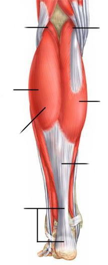
back muscles shins
The triceps muscle of the leg (m. triceps surae) has three heads. The gastrocnemius muscle (m. gastrocnemius) starts from the areas above the lateral and medial condyles of the thigh with two heads, forming the lower border of the fossa poplitea, and also, together with the posterior wall of the articular capsule, limits the entrance to the canalis cruropopliteus; the soleus muscle (m. soleus) is covered by the gastrocnemius muscle. Starting from the linea poplitea tibiae, the head of the fibula and the tendon arch stretched between the bones of the lower leg, it connects below into a single powerful calcaneal tendon of the triceps muscle of the lower leg - tendo calcaneus (Achillis), attached to the tuber of the calcaneus. Between the tendon and the calcaneal tuber there is a mucous bag.
Innervation: n. tibialis (LIV-SII).
Function. Flexes the foot at the ankle joint. When walking and running pushes the foot off the ground.
The plantar muscle (m. plantaris) starts from the area above the condyle of the thigh and the capsule of the knee joint. Then a thin tendon penetrates between the gastrocnemius and soleus muscles and is woven into the tendon of the triceps muscle of the lower leg.
Innervation and function. Same as the calf muscle.
The long flexor of the fingers (m. flexor digitorum longus) is located on the medial surface of the lower leg. It starts from the middle third of the posterior surface of the tibia and the deep fascia of the lower leg. The tendon reaches the medial malleolus and under the retinaculum mm. flexorum in the fibrous canal passes to the foot between the tendons m. tibialis posterior and m. flexor hallucis longus. On the foot it crosses with the tendon m. flexor hallucis longus, receiving from it a fibrous bundle of fibers. From the long flexor of the fingers also begins part of the muscle bundles m. quadratus plantae. Then the long flexor of the fingers is divided into four tendons, which, piercing the tendon of the short flexor of the fingers in the region of the phalanges, are attached to the base of the distal phalanges from II to V fingers.
Innervation: n. tibialis (LV-SI).
Function. Bends the fingers, on which the foot rests when walking, and the foot at the ankle joint.
The tibialis posterior muscle (m. tibialis posterior) (Fig. 199) starts from the interosseous membrane and the bones of the lower leg of the entire posterior surface. The lower part is covered by the flexors of the fingers. The squamous tendon runs behind the medial malleolus and inserts on the tuberosity of the navicular and all of the cuneiform bones.
Function. Flexes at the ankle joint and supinates the foot, participates in maintaining its arches.
199. Muscles of the lower leg, rear view.
1 - m. gastrocnemius; 2 - m. soleus; 3 - m. tibialis posterior; 4 - m. flexor hallucis longus; 5 - m. peroneus longus; 6 - m. peroneus brevis; 7 - m. flexor digitorum longus; 8 - m. popliteus
The long flexor of the first finger (m. flexor hallucis longus) is a more massive muscle than the long flexor of the fingers and the posterior tibial muscle. It is located lateral to the previous muscles, bordering on the long and short peroneal muscles. It starts from the fibula and the intermuscular septum. Passes behind the medial malleolus and sustentaculum tali, surrounded by the synovial sheath in the fibrous canal. Attached to the distal phalanx of the first finger. Sesamoid bones are often found in the tendon.
Innervation: n. tibialis (LV-SII).
Function. Bends I finger, supports the inner arch of the foot. Due to the fibrous bundle that has entered the long flexor of the fingers, to some extent it helps to bend the other fingers.
Among the muscles of the lower leg, the anterior, lateral and posterior muscle groups are distinguished. The anterior group mainly includes the extensors of the foot, the lateral group includes the flexors and pronators of the foot, and the posterior group includes the flexors and arch supports of the foot.
Rice. 135. Muscles of the lower leg (front view):
1 - long peroneal muscle; 2 - medial head calf muscle; 3 - anterior tibial muscle; 4 - soleus muscle; 5 - short peroneal muscle; 6 - long extensor of the fingers; 7 - upper extensor retinaculum; 8 - tendon of the anterior tibial muscle; 9 - lower extensor retinaculum
front group
The anterior tibialis muscle (m. tibialis anterior) (Fig. 90, 135, 142, 146) unbends and adducts the foot, raising its medial edge. A long, narrow, superficial muscle originating on the lateral condyle of the tibia and the interosseous membrane. The attachment site is located on the plantar surface of the medial sphenoid bone and on the base of the I metatarsal bone. There is also a dry bag of the anterior tibial muscle (bursa subtendinea m. tibialis anterioris).
The long extensor digitorum longus (Fig. 90, 135, 141, 142, 146) unbends II-V fingers, as well as the foot, raising its lateral (outer) edge along with the third peroneal muscle. The muscle starts from the upper epiphysis of the tibia, the head and anterior edge of the fibula and the interosseous membrane. The muscle passes into a long narrow tendon, which divides into five thin individual tendons. Four of them are attached to the back of the II-IV fingers in such a way that the middle bundles of tendons are attached to the base of the middle phalanx, and the lateral ones - to the base of the distal phalanx. The fifth tendon attaches to the base of the fifth metatarsal.
The long extensor of the thumb (m. extensor hallucis longus) (Fig. 136) unbends the thumb, as well as the foot itself, raising its medial edge. Partially covered by the two previous muscles, located between them. The point of its beginning is the lower part of the medial surface of the body of the fibula, and the attachment point is the base of the distal phalanx. Part of the tendon bundles fuses with the base of the proximal phalanx.
Lateral group
The long peroneal muscle (m. peroneus longus) (Fig. 135, 137, 138, 139, 144, 146) abducts and flexes the foot, lowering its medial edge. Located on the lateral surface of the leg. The muscle starts from the head and upper body of the fibula and is attached to the medial sphenoid bone and the base of the I-II metatarsal bones.
The short peroneal muscle (m. peroneus brevis) (Fig. 135, 136, 138, 139, 140) abducts and flexes the foot, raising its lateral edge. This long and thin muscle is located on the outer surface of the fibula. It is covered by the long peroneal muscle. The point of its beginning is located on the lower half of the lateral surface of the body of the fibula and the intermuscular septum. The place of attachment is the tuberosity of the V metatarsal bone.
back group
The back group includes two muscle groups.
Surface layer
The triceps muscle of the lower leg (m. triceps surae) flexes the lower leg at the knee joint, flexes and rotates the foot outward. With a fixed position of the foot, it pulls the lower leg and thigh backwards. The muscle consists of the superficial gastrocnemius muscle and the deep soleus muscle. Calf muscle (m. gastrocnemius) (Fig. 90, 132, 133, 134, 135, 137, 138, 146) has two heads. The medial head (caput mediale) starts from the medial epicondyle of the femur, and the lateral head (caput laterale) - from the lateral epicondyle. Both heads are connected into a common tendon and attached to the calcaneal tuber. The soleus muscle (m. soleus) (Fig. 90, 135, 137, 138, 139, 146) is covered by the gastrocnemius muscle, starts from the head and upper third of the posterior surface of the body of the fibula and from the line of the soleus muscle of the tibia. The muscle is attached on the calcaneal tubercle, growing together with the tendon of the gastrocnemius muscle. The common tendon in the lower third of the lower leg forms the calcaneal tendon (tendo calcaneus) (Fig. 137, 138), the so-called Achilles tendon. The mucous bag of the calcaneal tendon (bursa tendinis calcanei) is also located here.
The plantar muscle (m. plantaris) (Fig. 134, 137, 138) stretches the capsule of the knee joint during flexion and rotation of the lower leg. The muscle is rudimentary and unstable, has a spindle shape. Its point of origin is located on the lateral condyle of the femur and the bag of the knee joint, and the place of attachment is on the calcaneus.

Rice. 136. Muscles of the lower leg and foot (front view):
1 - articular muscle of the knee; 2 - square muscle of the thigh; 3 - short peroneal muscle; 4 - long extensor of the big toe; 5 - short extensor of the big toe; 6 - tendon of the long extensor of the big toe; 7 - short extensor of the fingers

Rice. 137. Muscles of the lower leg (back view):
1 - plantar muscle; 2 - gastrocnemius muscle: a) medial head, b) lateral head; 3 - soleus muscle; 4 - fascia of the lower leg; 5 - tendon of the posterior tibial muscle; 7 - tendon of the long flexor of the fingers; 8 - calcaneal tendon (Achilles tendon)

Rice. 138. Muscles of the lower leg (back view):
1 - plantar muscle; 2 - popliteal muscle; 3 - soleus muscle; 4 - tendon of the plantar muscle; 5 - gastrocnemius muscle: a) medial head, b) lateral head; 6 - tendon of the long peroneal muscle; 7 - tendon of the posterior tibial muscle; 8 - short peroneal muscle; 9 - tendon of the long flexor of the fingers; 10 - calcaneal tendon (Achilles tendon)

Rice. 139. Muscles of the lower leg (back view):
1 - popliteal muscle; 2 - soleus muscle; 4 - long peroneal muscle; 5 - long finger flexor; 6 - long flexor of the thumb; 7 - short peroneal muscle; 8 - flexor retainer; 9 - upper retainer of the long and short peroneal muscles

Rice. 140. Muscles of the lower leg and foot (rear view):
1 - popliteal muscle; 2 - short peroneal muscle; 3 - posterior tibial muscle; 4 - short flexor of the big toe; 5 - short flexor of the little toe of the foot; 6 - tendons of the long flexor of the fingers; 7 - interosseous muscles
deep layer
The popliteal muscle (m. popliteus) (Fig. 138, 139, 140) flexes the lower leg, rotating it inward and pulling the capsule of the knee joint. A short flat muscle, located on the posterior surface of the capsule of the knee joint, starts from it and from the lateral condyle of the femur, and is attached to the posterior surface of the body of the tibia.
The long flexor of the fingers (m. flexor digitorum longus) (Fig. 90, 137, 138, 139, 140, 143, 146) flexes the distal phalanges of the II-V fingers and takes part in the rotation of the foot outward, raising its medial edge. It is located on the posterior surface of the tibia, starts from the middle third of the posterior surface of the body of the tibia and from the deep sheet of the fascia of the leg. The tendon of the muscle is divided into four tendons, which are attached to the base of the distal phalanges of the II-V fingers.
The long flexor of the thumb (m. flexor hallucis longus) (Fig. 139, 143, 146) flexes the thumb, takes part in the flexion of the II-V fingers due to fibrous bundles, which are a continuation of the tendon, and also flexes and rotates the foot. The muscle originates from the lower two-thirds of the posterior surface of the body of the fibula and from the interosseous membrane, and is attached at the base of the distal phalanx of the thumb.
The tibialis posterior muscle (m. tibialis posterior) (Fig. 137, 138, 139, 140, 146) flexes and adducts the foot, rotating it outward. It is located on the interosseous membrane between the two previous muscles and is partially covered by the long flexor of the thumb. Its point of origin is on the posterior surfaces of the bodies of the tibia and fibula, and the place of attachment is on the sphenoid bones of the foot and the tuberosity of the scaphoid.


